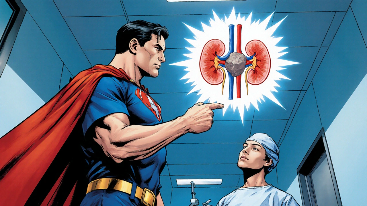
Kidney Stone Surgery: What to Expect and How to Prepare
A clear guide on what kidney stone surgery involves, how to prepare, what to expect on the day, recovery steps, risks, and practical tips for a smooth return to daily life.
When working with Percutaneous Nephrolithotomy, a minimally invasive surgical technique that removes kidney stones through a small skin incision. Also known as PCNL, it offers a safe alternative for treating large renal calculi and complex stone burdens. The method often pairs with laser lithotripsy to break stones into removable fragments, and it can be complemented by ureteroscopy when the anatomy demands a combined approach.
Percutaneous Nephrolithotomy is typically chosen when stones exceed 2 cm, when staghorn calculi fill the renal pelvis, or when shock wave therapy fails. The procedure starts with precise imaging—fluoroscopy or ultrasound guides the needle into the collecting system, establishing a tract that serves as a tunnel for the nephroscope. This imaging requirement creates a clear semantic link: PCNL requires real‑time visual guidance to ensure safe access. Once access is secured, the surgeon dilates the tract, inserts a working sheath, and uses a nephroscope to locate the stone.
The next step is stone fragmentation. Modern centers rely on holmium laser lithotripsy, which vaporizes stone material with minimal collateral damage. The laser’s precision reduces the need for larger access tracts, paving the way for mini‑PCNL or ultra‑mini approaches. After the stone is broken down, suction or a basket retrieves the fragments, and the tract is either closed with a nephrostomy tube or left tubeless for faster recovery. This sequence—access, dilation, lithotripsy, extraction—forms the backbone of PCNL and ties directly to the related entities of imaging, lasers, and endoscopic tools.
Recovery after PCNL is generally quicker than open surgery. Most patients spend one night in the hospital, and pain is manageable with standard analgesics. Potential complications include bleeding, infection, or injury to surrounding organs, but they occur in less than 5 % of cases when the procedure follows strict protocols. Follow‑up imaging confirms stone‑free status; if residual fragments remain, a secondary ureteroscopy can polish away leftovers without another full‑tract creation.
Advances keep reshaping the field. Mini‑PCNL uses tracts as small as 11–14 mm, reducing postoperative pain and hospital stay. The supine position, popularized in the last decade, allows simultaneous airway management and easier conversion to ureteroscopy if needed. Tubeless PCNL eliminates the nephrostomy tube altogether for selected patients, further speeding discharge. These innovations illustrate how PCNL evolves by integrating related concepts like tract size, patient positioning, and adjunctive endoscopic techniques.
Beyond the operating room, patient education plays a crucial role. Understanding when PCNL is appropriate, what the steps involve, and how postoperative care looks can empower patients to make informed choices. Nutrition, hydration, and metabolic evaluation help prevent recurrence, while regular follow‑up ensures any new stones are caught early.
Below you’ll find a curated set of articles that dive deeper into each aspect introduced here—comparisons of PCNL with other stone‑removal methods, detailed looks at laser settings, and guides on post‑procedure care. Use them to expand your knowledge, compare options, and plan the best treatment path for yourself or your patients.

A clear guide on what kidney stone surgery involves, how to prepare, what to expect on the day, recovery steps, risks, and practical tips for a smooth return to daily life.